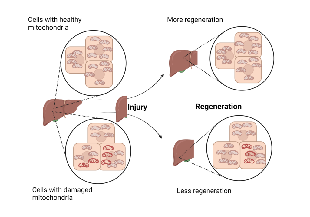
We all know that mitochondria are the powerhouse of cells because they generate energy from food. However, we did not know their involvement in cellular regeneration. Scientists at UT Southwestern Medical Center determined that liver cells with damaged mitochondria have less ability to regenerate when injured. Henceforth, allowing cells with undamaged mitochondria to grow more, rejuvenating the liver. This study was published in Science journal’s June 2024 issue.

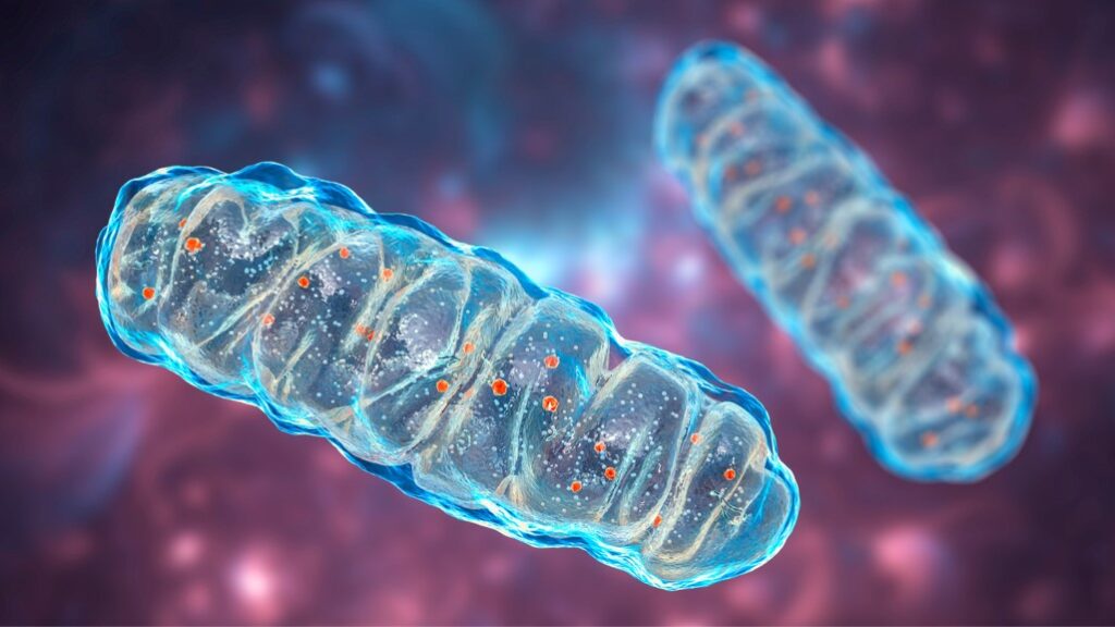
Image I: Stock Images
According to the CDC (Centers for Disease Control and Prevention), liver disease is at 10th rank for the cause of death in the United States. In human liver diseases– liver failure, cirrhosis, and fatty liver– the liver regeneration capacity is reduced but we do not know how. Past studies have shown that damaged mitochondria are present in liver cells in these diseases. This motivated the scientists to find out how damaged mitochondria could affect liver regeneration.
What did they do?
The scientists worked on mice to surgically remove 70% of their liver, forcing it to regenerate. They measured various metabolite levels in mitochondria to know which nutrients were used during regeneration. They observed lower fatty acid levels, due to increased fatty acid digestion of fats in the food. As a next step, they aimed to measure these levels in damaged mitochondria. To create damaged mitochondria, they mutated different genes of the electron transport chain (ETC). ETC is a pathway that generates energy in mitochondria. It is indirectly required for fatty acid digestion. Consequently, they observed reduced fatty acid digestion — or, increased fatty acid levels — that decreased liver regeneration. To confirm the role of fatty acids, they used chemical/genetic manipulation to increase fatty acid digestion in the damaged mitochondria. They found a subsequent increase in liver regeneration, hoping to use it as a potential treatment. Based on these findings they concluded that damaged mitochondria reduced liver regeneration by decreasing fatty acid digestion. Thereby allowing cells with healthy mitochondria to grow more, contributing to regeneration. This is a good example of natural selection at a cellular level to promote a healthy organ.

Image II: Mitochondria Rejuvenation
What are the next steps?
This study demonstrated that increasing fatty acid oxidation in damaged mitochondria improves mice’s liver regeneration ability. The next step would be to test these therapies in mouse liver disease models, which would mimic human disease. Other large animal models should also be used to validate these therapies. The ultimate test would be in clinical trials to know their efficacy and safety in humans. However, getting approval for clinical trials is a long process with multiple steps that could take several years.
How does this research affect humans?
In liver diseases, where most of the mitochondria are damaged, the liver is unable to rejuvenate. Therapies that increase fatty acid digestion can be used based on this study. However, patients with liver diseases have different underlying causes. These therapies could work only on those patients who have high fatty acid levels in their liver cells’ mitochondria. Thus, to determine who would respond to these drugs, patients’ biopsies need to be performed to measure mitochondria fatty acid levels. This study opens new directions to treat liver diseases, whose potential needs to be further verified.
Dr. Vinny Negi, Ph.D.
Sources
Wang X, Menezes CJ, Jia Y, et al. Metabolic inflexibility promotes mitochondrial health during liver regeneration. Science 2024 Jun 14;384(6701):eadj4301.doi: 10.1126/science.adj4301.
Disclaimer
The editors take care to share authentic information. In case of any discrepancies please write to newsletter@medness.org
The sponsors do not have any influence on the nature or kind of the news/analysis reported in MedNess. The views and opinions expressed in this article are those of the authors and do not necessarily reflect the official policy or position of MedNess. Examples of analysis performed within this article are only examples. They should not be utilized in real-world analytic products as they are based only on very limited and dated open-source information. Assumptions made within the analysis are not reflective of the position of anyone volunteering or working for MedNess. This blog is strictly for news and information. It does not provide medical advice, diagnosis or treatment nor investment suggestions. This content is not intended to be a substitute for professional medical advice, diagnosis, or treatment. Always seek the advice of your physician or another qualified health provider with any questions you may have regarding a medical condition. Never disregard professional medical advice or delay in seeking it because of something you have read on this website.
MedNess is a part of STEMPeers® which is a 501(c)(3) organization registered in PA as PhD Career Support Group. The organization helps create a growing network of STEM scientists that is involved in peer-to-peer mentoring and support.


